中文English
ISSN 1001-5256 (Print)
ISSN 2097-3497 (Online)
CN 22-1108/R
Volume 41 Issue 10
Oct.  2025
Turn off MathJax
Article Contents

Effect of tectorigenin on the viability, migration, and apoptosis of hepatoma cells and its mechanism

DOI: 10.12449/JCH251019
Research funding:

The Basic Research Program of Guizhou Provincial Science and Technology Department (Qiankehe Foundation-ZK (2022) General 452);

National Natural Science Foundation of China Regional Funds (82160119);

National Natural Science Foundation of China Regional Funds (82360122)

More Information
  • Corresponding author: ZHU Juanjuan, 184184239@qq.com (ORCID: 0009-0002-4999-4248)
  • Received Date: 2025-03-24
  • Accepted Date: 2025-04-14
  • Published Date: 2025-10-25
  •   Objective  To investigate the effect of blueberry-derived tectorigenin (TEC) on hepatocellular carcinoma cell lines HepG2 and Huh7 and its mechanism.  Methods  TEC was extracted from blueberries and purified, and a bioinformatics analysis was performed to identify potential target genes and signaling pathways. HepG2 and Huh7 cell lines were used and divided into 0, 30, 60, and 90 μg/mL groups according to the concentration of TEC. CCK-8 assay was used to measure cell viability; wound healing assay and Transwell assay were used to assess the migration ability of cells; flow cytometry was used to measure cell apoptosis rate; Western Blot was used to measure the protein expression levels of CCNB1, p53, MDM2, Bax, Bcl-2, and active-Caspase 3. Cell models with low CCNB1 expression (NC group, si-NC group, si-NC+TEC group, si-CCNB1 group, and si-CCNB1+TEC group) and CCNB1 overexpression (OE-NC group, OE-NC+TEC group, OE-CCNB1 group, and OE-CCNB1+TEC group) were established to validate the targets. A one-way analysis of variance or two factors analysis of variance was used for comparison of continuous data between multiple groups, and the least significant difference t-test was used for further comparison between two groups. The Wilcoxon signed rank sum test was used to compare the expression levels of genes between cancer tissue and paracancerous tissue.  Results  In HepG2 and Huh7 cells under the same concentration of TEC, cell viability at 24 hours of TEC intervention was significantly lower than that at 12 and 48 hours (all P<0.05), and at 24 hours of intervention, the TEC 90 μg/mL group had a significantly lower cell viability than the other groups (all P<0.05). Therefore, TEC intervention for 24 hours at a concentration of 90 μg/mL was used for subsequent studies. Compared with the TEC 0 μg/mL group, the 30, 60, and 90 μg/mL groups had significant reductions in the number of migrated cells and wound healing rate (all P<0.05), and compared with the NC group and si-NC group, the si-NC+TEC group and the si-CCNB1 group for HepG2 and Huh7 cells had significant reductions in the number of migrated cells and wound healing rate (all P<0.05). Compared with the NC group and si-NC group, the si-NC+TEC group and the si-CCNB1 group for HepG2 and Huh7 cells had a significant increase in cell apoptosis rate (all P<0.05). For HepG2 cells, compared with the 0 μg/mL group, the 30, 60, and 90 μg/mL groups had significant reductions in the protein expression levels of CCNB1 and Bcl-2 (all P<0.05), and the 60 and 90 μg/mL groups had significant increases in the protein expression levels of p53, Bax, and active-Caspase 3 (all P<0.001) and a significant reduction in the protein expression level of MDM2 (both P<0.05). For Huh7 cells, compared with the 0 μg/mL group, the 30, 60, and 90 μg/mL groups had a significant reduction in the protein expression level of CCNB1 (all P<0.01); the 60 and 90 μg/mL groups had significant increases in the protein expression levels of p53 and Bax and a significant reduction in the protein expression level of MDM2 (all P<0.05); the 90 μg/mL group had a significant reduction in the protein expression level of Bcl-2 and a significant increase in the protein expression level of active-Caspase 3 (both P<0.01). Compared with the si-NC group, the si-NC+TEC group and the si-CCNB1 group for HepG2 and Huh7 cells had significant reductions in the protein expression levels of CCNB1, MDM2, and Bcl-2 and significant increases in the protein expression levels of p53 and Bax (all P<0.05). Compared with the OE-NC group, the OE-NC+TEC group for HepG2 and Huh7 cells had significant reductions in the protein expression levels of CCNB1 and MDM2 and a significant increase in the protein expression level of p53 (all P<0.05), while the OE-CCNB1 group had significant increases in the protein expression levels of CCNB1 and MDM2 and a significant reduction in the protein expression level of p53 (all P<0.05), and there were no significant differences in the protein expression level of CCNB1, MDM2, and p53 between the OE-CCNB1 group and the OE-CCNB1+TEC group (all P>0.05).  Conclusion  TEC can inhibit the proliferation and migration of HepG2 and Huh7 cells and promote their apoptosis in vitro, possibly by downregulating the expression of CCNB1 and activating the p53 signaling pathway.


  • loading
  • [1]
    TOH MR, WONG EYT, WONG SH, et al. Global epidemiology and genetics of hepatocellular carcinoma[J]. Gastroenterology, 2023, 164( 5): 766- 782. DOI: 10.1053/j.gastro.2023.01.033.
    [2]
    SILVA S, COSTA EM, VEIGA M, et al. Health promoting properties of blueberries: A review[J]. Crit Rev Food Sci Nutr, 2020, 60( 2): 181- 200. DOI: 10.1080/10408398.2018.1518895.
    [3]
    ZHAN W, LIAO X, YU L, et al. Effects of blueberries on migration, invasion, proliferation, the cell cycle and apoptosis in hepatocellular carcinoma cells[J]. Biomed Rep, 2016, 5( 5): 579- 584. DOI: 10.3892/br.2016.774.
    [4]
    RONG J, FU F, HAN CX, et al. Tectorigenin: A review of its sources, pharmacology, toxicity, and pharmacokinetics[J]. Molecules, 2023, 28( 15): 5904. DOI: 10.3390/molecules28155904.
    [5]
    JIANG CP, DING H, SHI DH, et al. Pro-apoptotic effects of tectorigenin on human hepatocellular carcinoma HepG2 cells[J]. World J Gastroenterol, 2012, 18( 15): 1753- 1764. DOI: 10.3748/wjg.v18.i15.1753.
    [6]
    YEH LT, HSU LS, CHUNG YH, et al. Tectorigenin inhibits glioblastoma proliferation by G0/G1 cell cycle arrest[J]. Medicina(Kaunas), 2020, 56( 12): 681. DOI: 10.3390/medicina56120681.
    [7]
    ZENG LW, YUAN SF, SHEN JL, et al. Suppression of human breast cancer cells by tectorigenin through downregulation of matrix metalloproteinases and MAPK signaling in vitro[J]. Mol Med Rep, 2018, 17( 3): 3935- 3943. DOI: 10.3892/mmr.2017.8313.
    [8]
    FANG R, HOUGHTON PJ, HYLANDS PJ. Cytotoxic effects of compounds from Iris tectorum on human cancer cell lines[J]. J Ethnopharmacol, 2008, 118( 2): 257- 263. DOI: 10.1016/j.jep.2008.04.006.
    [9]
    YANG YJ, KE TY, LIU SX, et al. Synergistic sensitization of apatinib mesylate and radiotherapy on hepatocarcinoma cells in vitro[J]. J Jilin Univ(Med Edit), 2024, 50( 4): 1009- 1015. DOI: 10.13481/j.1671-587X.2024-04015.

    杨永净, 柯天洋, 刘士新, 等. 甲磺酸阿帕替尼联合放疗对肝癌HepG2细胞的体外协同增敏作用[J]. 吉林大学学报(医学版), 2024, 50( 4): 1009- 1015. DOI: 10.13481/j.1671-587X.202404015.
    [10]
    NAEEM A, HU PY, YANG M, et al. Natural products as anticancer agents: Current status and future perspectives[J]. Molecules, 2022, 27( 23): 8367. DOI: 10.3390/molecules27238367.
    [11]
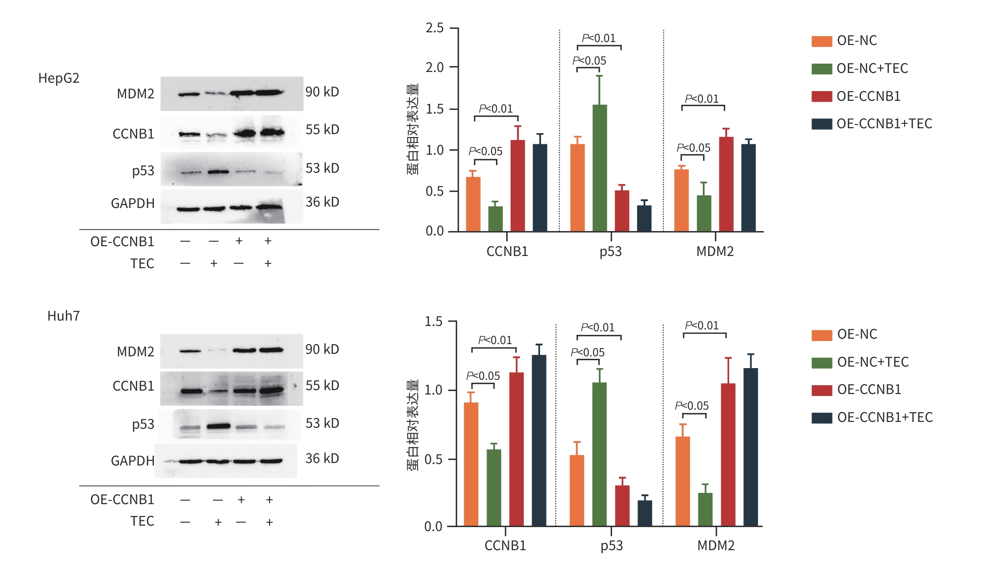
    ZHAN G, PAN LQ, TU K, et al. Antitumor, antioxidant, and nitrite scavenging effects of Chinese water chestnut(Eleocharis dulcis) peel flavonoids[J]. J Food Sci, 2016, 81( 10): H2578- H2586. DOI: 10.1111/1750-3841.13434.
    [12]
    KIM EM, JUNG CH, KIM J, et al. The p53/p21 complex regulates cancer cell invasion and apoptosis by targeting bcl-2 family proteins[J]. Cancer Res, 2017, 77( 11): 3092- 3100. DOI: 10.1158/0008-5472.CAN-16-2098.
    [13]
    GAVET O, PINES J. Progressive activation of CyclinB1-Cdk1 coordinates entry to mitosis[J]. Dev Cell, 2010, 18( 4): 533- 543. DOI: 10.1016/j.devcel.2010.02.013.
    [14]
    FANG YF, YU H, LIANG X, et al. Chk1-induced CCNB1 overexpression promotes cell proliferation and tumor growth in human colorectal cancer[J]. Cancer Biol Ther, 2014, 15( 9): 1268- 1279. DOI: 10.4161/cbt.29691.
    [15]
    LUNDGREN C, AHLIN C, HOLMBERG L, et al. Cyclin E1 is a strong prognostic marker for death from lymph node negative breast cancer. A population-based case-control study[J]. Acta Oncol, 2015, 54( 4): 538- 544. DOI: 10.3109/0284186X.2014.965274.
    [16]
    ZHOU L, LI J, ZHAO YP, et al. The prognostic value of Cyclin B1 in pancreatic cancer[J]. Med Oncol, 2014, 31( 9): 107. DOI: 10.1007/s12032-014-0107-4.
    [17]
    ZOU YP, RUAN SY, JIN L, et al. CDK1, CCNB1, and CCNB2 are prognostic biomarkers and correlated with immune infiltration in hepatocellular carcinoma[J]. Med Sci Monit, 2020, 26: e925289. DOI: 10.12659/MSM.925289.
    [18]
    WANG HL, GUO M, WEI HD, et al. Targeting p53 pathways: Mechanisms, structures, and advances in therapy[J]. Signal Transduct Target Ther, 2023, 8( 1): 92. DOI: 10.1038/s41392-023-01347-1.
    [19]
    KOO N, SHARMA AK, NARAYAN S. Therapeutics targeting p53-MDM2 interaction to induce cancer cell death[J]. Int J Mol Sci, 2022, 23( 9): 5005. DOI: 10.3390/ijms23095005.
    [20]
    LOU J, ZHAO L, ZHU YJ, et al. Effect of Fuzheng Ruanjian Anticancer Formula on malignant biological behaviors of hepatocellulars carcinoma HepG2 cells by regulating Akt/MDM2/P53 signaling pathway[J]. J Jilin Univ(Med Edit), 2024, 50( 6): 1654- 1663. DOI: 10.13481/j.1671-587X.20-240619.

    娄静, 赵雷, 朱岩洁, 等. 扶正软坚抗癌方调控Akt/MDM2/P53信号通路对肝癌HepG2细胞恶性生物学行为的影响[J]. 吉林大学学报(医学版), 2024, 50( 6): 1654- 1663. DOI: 10.13481/j.1671-587X.20240619.
    [21]
    YUAN JP, YAN RL, KRÄMER A, et al. Cyclin B1 depletion inhibits proliferation and induces apoptosis in human tumor cells[J]. Oncogene, 2004, 23( 34): 5843- 5852. DOI: 10.1038/sj.onc.1207757.
    [22]
    ZHANG H, ZHANG X, LI X, et al. Effect of CCNB1 silencing on cell cycle, senescence, and apoptosis through the p53 signaling pathway in pancreatic cancer[J]. J Cell Physiol, 2018, 234( 1): 619- 631. DOI: 10.1002/jcp.26816.
    [23]
    XIA P, ZHANG H, XU KQ, et al. MYC-targeted WDR4 promotes proliferation, metastasis, and sorafenib resistance by inducing CCNB1 translation in hepatocellular carcinoma[J]. Cell Death Dis, 2021, 12( 7): 691. DOI: 10.1038/s41419-021-03973-5.
  • 加载中

Catalog

    通讯作者: 陈斌, bchen63@163.com
    • 1. 

      沈阳化工大学材料科学与工程学院 沈阳 110142

    1. 本站搜索
    2. 百度学术搜索
    3. 万方数据库搜索
    4. CNKI搜索

    Figures(10)  / Tables(1)

    Article Metrics

    Article views (46) PDF downloads(12) Cited by()
    Proportional views
    Related

    /

    DownLoad:  Full-Size Img  PowerPoint
    Return
    Return