

 PDF下载 ( 1251 KB)

T细胞衰老在老年慢性乙型肝炎患者肝纤维化向肝硬化进展中的作用机制
DOI: 10.12449/JCH260835
利益冲突声明:本文不存在任何利益冲突。
作者贡献声明:王婷负责综述的总体设计,梳理文献及撰写初稿;杨成凯负责机制分析部分的内容深化;徐栩负责全文的学术审校,观点整合并最终定稿;王婷、杨成凯和徐栩共同完成资料核查与文稿修订。
Mechanism of action of T cell senescence in the progression of liver fibrosis to liver cirrhosis in elderly patients with chronic hepatitis B
-
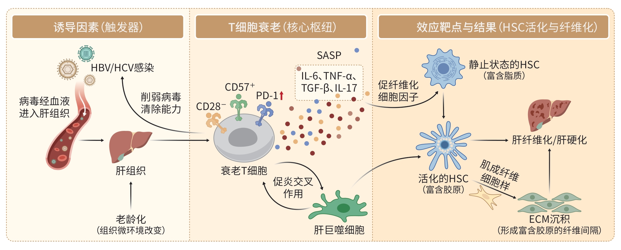
摘要: 肝纤维化是慢性肝病向肝硬化转变的关键病理过程,尤其在老年慢性乙型肝炎患者中,该过程进展迅速且预后较差。随着年龄增长,T细胞功能逐渐衰退,T细胞衰老被认为是加速肝纤维化进展的重要免疫学机制。当前研究表明,衰老T细胞通过改变肝脏炎症微环境、调节纤维化相关细胞活性及削弱免疫监视功能,促进肝纤维化向肝硬化的转化。本文系统综述了T细胞衰老的分子机制及其在老年慢性乙型肝炎肝纤维化进展中的作用,结合最新免疫学发现及干细胞治疗策略,探讨靶向免疫衰老的新型治疗途径,旨在为此类患者的精准干预提供理论支持。Abstract: Liver fibrosis is a key pathological process in the transition from chronic liver disease to cirrhosis, especially in elderly patients with chronic hepatitis B, and this process is characterized by rapid progression and poor prognosis. T cell function gradually declines with aging, and T cell senescence is recognized as a key immunological mechanism accelerating the progression of liver fibrosis. Current studies have shown that senescent T cells promote the transition from liver fibrosis to liver cirrhosis by altering the liver inflammatory microenvironment, modulating the activity of fibrosis-related cells, and impairing immune surveillance function. This article systematically reviews the molecular mechanisms of T cell senescence and its role in the progression of liver fibrosis in elderly patients with chronic hepatitis B, as well as novel therapeutic approaches targeting immunosenescence with reference to the latest immunological findings and stem cell-based therapeutic strategies, so as to provide theoretical support for precise intervention for such patients.
-
Key words:
- Hepatitis B, Chronic /
- Hepatic Fibrosis /
- Liver Cirrhosis /
- T-Lymphocytes /
- Cellular Senescence
-
[1] Jia Y H, Ma L Y, Wang Y D, et al. NLRP3 inflammasome and related cytokines reflect the immune status of patients with HBV-ACLF[J]. Mol Immunol, 2020, 120: 179- 186. DOI: 10.1016/j.molimm.2020.01.011. [2] Zecca A, Barili V, Canetti D, et al. Energy metabolism and cell motility defect in NK-cells from patients with hepatocellular carcinoma[J]. Cancer Immunol Immunother, 2020, 69( 8): 1589- 1603. DOI: 10.1007/s00262-020-02561-4. [3] Chen Q B, Lai L M, Chi X L, et al. CD19+CD24hiCD38hi B cell dysfunction in primary biliary cholangitis[J]. Mediators Inflamm, 2020, 2020: 3019378. DOI: 10.1155/2020/3019378. [4] Yang X X, Li J W, Ren M L, et al. Comprehensive analysis of immune signatures in primary biliary cholangitis and autoimmune hepatitis[J]. J Leukoc Biol, 2024, 117( 1): qiae085. DOI: 10.1093/jleuko/qiae085. [5] Li B, Xiong W Y, Zuo W, et al. Proximal telomeric decompaction due to telomere shortening drives FOXC1-dependent myocardial senescence[J]. Nucleic Acids Res, 2024, 52( 11): 6269- 6284. DOI: 10.1093/nar/gkae274. [6] Wang M, Guo Z, Zhao S S, et al. CD49d promotes T-cell senescence in chronic lymphocytic leukaemia[J]. Br J Haematol, 2025, 207( 1): 80- 91. DOI: 10.1111/bjh.20135. [7] Pangrazzi L, Weinberger B. T cells, aging and senescence[J]. Exp Gerontol, 2020, 134: 110887. DOI: 10.1016/j.exger.2020.110887. [8] Slaets H, Veeningen N, de Keizer P L J, et al. Are immunosenescent T cells really senescent[J]. Aging Cell, 2024, 23( 10): e14300. DOI: 10.1111/acel.14300. [9] Kasamatsu T. Implications of senescent T cells for cancer immunotherapy[J]. Cancers, 2023, 15( 24): 5835. DOI: 10.3390/cancers15245835. [10] Nakagami H. Cellular senescence and senescence-associated T cells as a potential therapeutic target[J]. Geriatr Gerontol Int, 2020, 20( 2): 97- 100. DOI: 10.1111/ggi.13851. [11] Nga H T, Nguyen T L, Yi H S. T-cell senescence in human metabolic diseases[J]. Diabetes Metab J, 2024, 48( 5): 864- 881. DOI: 10.4093/dmj.2024.0140. [12] Laphanuwat P, Gomes D C O, Akbar A N. Senescent T cells: Beneficial and detrimental roles[J]. Immunol Rev, 2023, 316( 1): 160- 175. DOI: 10.1111/imr.13206. [13] Huang L, Zhang C G, Jiang A M, et al. T-cell senescence in the tumor microenvironment[J]. Cancer Immunol Res, 2025, 13( 5): 618- 632. DOI: 10.1158/2326-6066.CIR-24-0894. [14] Mathiasen S L, Gall-Mas L, Pateras I S, et al. Bacterial genotoxins induce T cell senescence[J]. Cell Rep, 2021, 35( 10): 109220. DOI: 10.1016/j.celrep.2021.109220. [15] Fukushima Y, Ueno R, Minato N, et al. Senescence-associated T cells in immunosenescence and diseases[J]. Int Immunol, 2025, 37( 3): 143- 152. DOI: 10.1093/intimm/dxae056. [16] Covre L P, de Maeyer R P H, Gomes D C O, et al. The role of senescent T cells in immunopathology[J]. Aging Cell, 2020, 19( 12): e13272. DOI: 10.1111/acel.13272. [17] Yi H S, Kim S Y, Kim J T, et al. T-cell senescence contributes to abnormal glucose homeostasis in humans and mice[J]. Cell Death Dis, 2019, 10( 3): 249. DOI: 10.1038/s41419-019-1494-4. [18] Sim B C, Kang Y E, You S K, et al. Hepatic T-cell senescence and exhaustion are implicated in the progression of fatty liver disease in patients with type 2 diabetes and mouse model with nonalcoholic steatohepatitis[J]. Cell Death Dis, 2023, 14( 9): 618. DOI: 10.1038/s41419-023-06146-8. [19] Shive C L, Kowal C M, Desotelle A F, et al. Endotoxemia associated with liver disease correlates with systemic inflammation and T cell exhaustion in hepatitis C virus infection[J]. Cells, 2023, 12( 16): 2034. DOI: 10.3390/cells12162034. [20] Zhang H Y, Wang X C, Zhang X, et al. Hepatic ferroptosis induced by Clonorchis sinensis exacerbates liver fibrosis[J]. PLoS Negl Trop Dis, 2025, 19( 6): e0013164. DOI: 10.1371/journal.pntd.0013164. [21] Yashaswini C N, Qin T Y, Bhattacharya D, et al. Phenotypes and ontogeny of senescent hepatic stellate cells in metabolic dysfunction-associated steatohepatitis[J]. J Hepatol, 2024, 81( 2): 207- 217. DOI: 10.1016/j.jhep.2024.03.014. [22] Ezhilarasan D. Hepatic stellate cells in the injured liver: Perspectives beyond hepatic fibrosis[J]. J Cell Physiol, 2022, 237( 1): 436- 449. DOI: 10.1002/jcp.30582. [23] Shrivastava S, Kottilil S, Sherman K E, et al. CCR5+ T-cells homed to the liver exhibit inflammatory and profibrogenic signatures in chronic HIV/HCV-coinfected patients[J]. Viruses, 2021, 13( 10): 2074. DOI: 10.3390/v13102074. [24] Wu J Z, Zhang L, Zhao Z H, et al. Advancing T-cell immunotherapy for cellular senescence and disease: Mechanisms, challenges, and clinical prospects[J]. Ageing Res Rev, 2025, 109: 102783. DOI: 10.1016/j.arr.2025.102783. [25] Chai J Q, Li L Y, Wu Q M, et al. CD96: Immunoregulation and its role and prospect in immunotherapy of primary hepatocellular carcinoma[J]. Eur J Gastroenterol Hepatol, 2025, 37( 5): 534- 539. DOI: 10.1097/MEG.0000000000002916. [26] Zhang Q S, Wang J N, Yang T L, et al. SHMT2 regulates CD8+ T cell senescence via the reactive oxygen species axis in HIV-1 infected patients on antiretroviral therapy[J]. eBioMedicine, 2025, 112: 105533. DOI: 10.1016/j.ebiom.2024.105533. [27] Turano P S, Akbulut E, Dewald H K, et al. Epigenetic mechanisms regulating CD8+ T cell senescence in aging humans[J]. bioRxiv, 2025. DOI: 10.1101/2025.01.17.633634.[ Epub ahead of print] [28] Wang S, Wang X O, Zheng X Y, et al. NGR-modified curcumin nanovesicles reverse immunotherapy resistance in triple-negative breast cancer via TLR9 and mTOR pathway modulation[J]. Cell Biol Toxicol, 2025, 41( 1): 109. DOI: 10.1007/s10565-025-10055-1. [29] Admasu T D, Yu J S. Harnessing immune rejuvenation: Advances in overcoming T cell senescence and exhaustion in cancer immunotherapy[J]. Aging Cell, 2025, 24( 5): e70055. DOI: 10.1111/acel.70055. [30] Khosla S, Farr J N, Tchkonia T, et al. The role of cellular senescence in ageing and endocrine disease[J]. Nat Rev Endocrinol, 2020, 16( 5): 263- 275. DOI: 10.1038/s41574-020-0335-y. [31] Nazarie Ignat S R, Gharbia S, Hermenean A, et al. Regenerative potential of mesenchymal stem cells’(MSCs) secretome for liver fibrosis therapies[J]. Int J Mol Sci, 2021, 22( 24): 13292. DOI: 10.3390/ijms222413292. [32] Bozorgi V, Babaahmadi M, Salehi M, et al. From signaling pathways to clinical trials: Mesenchymal stem cells as multimodal regenerative architects in liver cirrhosis therapy[J]. Stem Cell Res Ther, 2025, 16( 1): 421. DOI: 10.1186/s13287-025-04535-8. [33] Janelle V, Delisle J S. T-cell dysfunction as a limitation of adoptive immunotherapy: Current concepts and mitigation strategies[J]. Cancers, 2021, 13( 4): 598. DOI: 10.3390/cancers13040598. [34] Calviño C, Ceballos C, Alfonso A, et al. Optimization of universal allogeneic CAR-T cells combining CRISPR and transposon-based technologies for treatment of acute myeloid leukemia[J]. Front Immunol, 2023, 14: 1270843. DOI: 10.3389/fimmu.2023.1270843. [35] Liu Q W, Li J F, Sun X Q, et al. Immunosenescence and cancer: Molecular hallmarks, tumor microenvironment remodeling, and age-specific immunotherapy challenges[J]. J Hematol Oncol, 2025, 18( 1): 81. DOI: 10.1186/s13045-025-01735-w. [36] Li Y K, Yi J S, Howard J F, et al. Cellular changes in eculizumab early responders with generalized myasthenia gravis[J]. Clin Immunol, 2021, 231: 108830. DOI: 10.1016/j.clim.2021.108830. [37] Takasugi M, Yoshida Y, Ohtani N. Cellular senescence and the tumour microenvironment[J]. Mol Oncol, 2022, 16( 18): 3333- 3351. DOI: 10.1002/1878-0261.13268. [38] Wang X D, Wang J, Peng H D, et al. Role of immune cell interactions in alcohol-associated liver diseases[J]. Liver Res, 2024, 8( 2): 72- 82. DOI: 10.1016/j.livres.2024.06.002. [39] Zeng W F, Liu F R, Liu Y C, et al. Targeting TM4SF1 promotes tumor senescence enhancing CD8+ T cell cytotoxic function in hepatocellular carcinoma[J]. Clin Mol Hepatol, 2025, 31( 2): 489- 508. DOI: 10.3350/cmh.2024.0934. [40] Sakai Y, Fukunishi S, Takamura M, et al. Clinical trial of autologous adipose tissue-derived regenerative(stem) cells therapy for exploration of its safety and efficacy[J]. Regen Ther, 2021, 18: 97- 101. DOI: 10.1016/j.reth.2021.04.003. [41] Sakai Y, Fukunishi S, Takamura M, et al. Regenerative therapy for liver cirrhosis based on intrahepatic arterial infusion of autologous subcutaneous adipose tissue-derived regenerative(stem) cells: Protocol for a confirmatory multicenter uncontrolled clinical trial[J]. JMIR Res Protoc, 2020, 9( 3): e17904. DOI: 10.2196/17904. -

 下载:
下载:
 本文二维码
本文二维码
图(1)
计量
- 文章访问数: 8
- HTML全文浏览量: 5
- PDF下载量: 1
- 被引次数: 0


