

 PDF下载 ( 22102 KB)

鳖甲煎丸对四氯化碳诱导的肝纤维化大鼠模型的影响及其作用机制
DOI: 10.12449/JCH260819
Effect and mechanism of action of Biejiajian Pills on a rat model of hepatic fibrosis induced by carbon tetrachloride
-
摘要:
目的 探讨鳖甲煎丸对四氯化碳诱导的大鼠肝纤维化的治疗作用及其基于胆汁酸-短链脂肪酸代谢轴的机制。 方法 采用四氯化碳诱导的大鼠肝纤维化模型,将32只SD雄性大鼠随机分为对照组、模型组、鳖甲煎丸组(2.2 g/kg)和水飞蓟素组(43.19 mg/kg),每组8只。除对照组外,其余各组腹腔注射四氯化碳(4∶6玉米油稀释,2 mL/kg)诱导肝纤维化模型,连续8周,给药组同时灌胃相应药物。采用全自动生化分析仪检测血清丙氨酸氨基转移酶(ALT)、天冬氨酸氨基转移酶(AST)、总胆红素(TBil)和白蛋白(Alb)水平;氯胺T比色法检测肝组织羟脯氨酸(HYP)含量;苏木精-伊红染色和马松染色观察肝组织病理学变化;UPLC-MS/MS和GC-MS分别检测胆汁酸和短链脂肪酸水平。所有数据均经Shapiro-Wilk检验和Levene检验进行正态性和方差齐性验证;满足正态分布的计量资料采用单因素方差分析进行多组间比较,进一步两两比较采用LSD-t检验;Ishak评分多组间比较采用Kruskal-Wallis H检验,进一步两两比较采用Dunn’s检验。对代谢组学数据中有差异的代谢物进行筛选,采用Benjamini-Hochberg法进行多重比较校正。结合正交偏最小二乘判别分析(OPLS-DA)模型得到变量重要性投影(VIP)值,选取VIP>1且P<0.05的代谢物作为组间的显著差异代谢物。OPLS-DA模型的稳健性通过200次置换检验进行评估,并报告模型的主要参数模型,包括对分类变量的解释率(R2Y)和模型的预测能力(Q2)。采用Spearman进行等级相关性分析。 结果 染色结果显示,鳖甲煎丸组肝细胞排列较整齐,炎性细胞浸润和胶原纤维沉积明显减轻。与对照组相比,模型组血清TBil和HYP水平均显著升高,Alb水平显著降低(P值均<0.05)。与对照组相比,鳖甲煎丸组血清AST、ALT、TBil和HYP水平显著升高,Alb水平显著降低(P值均<0.05);水飞蓟素组血清ALT、TBil和HYP水平显著升高,Alb水平显著降低(P值均<0.05)。与模型组比较,鳖甲煎丸组血清ALT、AST、TBil和HYP水平均显著降低(P值均<0.05),水飞蓟素组血清ALT、TBil、HYP和AST水平显著降低,Alb水平显著升高(P值均<0.05)。机制研究表明,与对照组相比,模型组牛磺石胆酸、牛磺-α-鼠胆酸、牛磺胆酸、甘氨脱氧胆酸、牛磺猪胆酸、鹅去氧胆酸、甘氨猪去氧胆酸、7,12-二酮石胆酸、牛磺鹅去氧胆酸和甘氨鹅去氧胆酸水平均显著下降(P值均<0.05),猪去氧胆酸、去甲胆酸、β-鼠胆酸和胆酸水平均显著提高(P值均<0.05)。与模型组比较,鳖甲煎丸组保护性胆汁酸牛磺石胆酸、牛磺胆酸、牛磺猪胆酸、鹅去氧胆酸和牛磺脱氧胆酸水平均显著升高(P值均<0.05),牛磺-α-鼠胆酸、牛磺-β-鼠胆酸、牛磺猪去氧胆酸+牛磺熊去氧胆酸、α-鼠胆酸、甘氨熊去氧胆酸和去甲胆酸水平均显著降低(P值均<0.05);短链脂肪酸中乙酸、丙酸、异丁酸、丁酸、异戊酸、戊酸和己酸水平均显著升高(P值均<0.05)。与模型组相比,水飞蓟素组牛磺石胆酸、牛磺-α-鼠胆酸、去甲胆酸、甘氨胆酸、脱氧胆酸、牛磺-β-鼠胆酸、牛磺脱氧胆酸、牛磺猪去氧胆酸+牛磺熊去氧胆酸和α-鼠胆酸水平均显著下降(P值均<0.05),牛磺胆酸和鹅去氧胆酸显著升高(P值均<0.05),短链脂肪酸中乙酸、丙酸、戊酸和己酸水平均显著升高(P值均<0.05)。相关性分析结果表示, 胆汁酸及短链脂肪酸代谢与血清指标的变化之间存在密切联系。 结论 鳖甲煎丸可通过调节胆汁酸-短链脂肪酸代谢轴改善四氯化碳诱导的肝纤维化大鼠模型。 -
关键词:
- 肝纤维化 /
- 鳖甲煎丸 /
- 水飞蓟素 /
- 大鼠, Sprague-Dawley
Abstract:Objective To investigate the therapeutic effect of Biejiajian Pills on rats with carbon tetrachloride (CCl4)-induced hepatic fibrosis and its mechanism based on the bile acid (BA)-short-chain fatty acid (SCFA) metabolic axis. Methods The method of CCl4 induction was used to establish a rat model of hepatic fibrosis, and 32 male Sprague-Dawley rats were randomly divided into control group, model group, Biejiajian Pills group (2.2 g/kg), and silymarin group (43.19 mg/kg), with 8 rats in each group. All rats except those in the control group were given intraperitoneal injection of CCl4 (diluted with corn oil at a ratio of 4∶6, 2 mL/kg) to induce a model of hepatic fibrosis for 8 consecutive weeks, and the rats in the drug administration groups were given the corresponding drug by gavage. An automatic biochemical analyzer was used to measure the serum levels of alanine aminotransferase (ALT), aspartate aminotransferase (AST), total bilirubin (TBil), and albumin (Alb); chloramine-T colorimetry was used to measure the content of hydroxyproline (HYP) in liver tissue; HE staining and Masson staining were used to observe liver histopathological changes; UPLC-MS/MS and GC-MS were used to measure the levels of BAs and SCFAs. The Shapiro-Wilk test and the Levene test were performed for all data to determine normality and homogeneity of variance; a one-way analysis of variance was used for comparison of normally distributed continuous data between multiple groups, and the least significant difference t-test was used for further comparison between two groups; the Kruskal-Wallis H test was used for comparison of Ishak score between groups, and the Dunn’s test was used for further comparison between two groups. The Benjamini-Hochberg method was used for multiple comparison correction in the screening of differentially expressed metabolites in metabolomics data. The variable importance in projection (VIP) values were obtained in combination with the orthogonal partial least squares-discriminant analysis (OPLS-DA) model, and metabolites with VIP>1 and P<0.05 were selected as significantly differentially expressed metabolites between groups. The robustness of the OPLS-DA model was evaluated through 200 permutation tests, and the main parameters of the model were reported, including the interpretation rate of the model for categorical variables (R2Y) and the predictive ability of the model (Q2). A Spearman’s rank correlation analysis was performed. Results The results of staining showed ordered arrangement of hepatocytes in the Biejiajian Pills group, with significant alleviation of inflammatory cell infiltration and collagen fiber deposition. Compared with the control group, the model group had significant increases in the serum levels of TBil and HYP and a significant reduction in the serum level of Alb (all P<0.05). Compared with the control group, the Biejiajian Pills group had significant increases in the serum levels of AST, ALT, TBil, and HYP and a significant reduction in the serum level of Alb (all P<0.05), and the silymarin group had significant increases in the serum levels of ALT, TBil, and HYP and a significant reduction in the serum level of Alb (all P<0.05). Compared with the model group, the Biejiajian Pills group had significant reductions in the serum levels of ALT, AST, TBil, and HYP (all P<0.05), and the silymarin group had significant reductions in the serum levels of ALT, TBil, HYP, and AST and a significant increase in the serum level of Alb (all P<0.05). Mechanism studies showed that compared with the control group, the model group had significant reductions in the levels of taurolithocholic acid, tauro-α-muricholic acid, taurocholic acid, glycodeoxycholic acid, taurohyocholic acid, chenodeoxycholic acid, glycohyodeoxycholic acid, 7,12-diketolithocholic acid, taurochenodeoxycholic acid, and glycochenodeoxycholic acid and significant increases in the levels of hyodeoxycholic acid, norcholic acid, β-muricholic acid, and cholic acid (all P<0.05). Compared with the model group, the Biejiajian Pills group had significant increases in the levels of the protective BAs taurolithocholic acid, chenodeoxycholic acid, taurocholic acid, taurohyocholic acid, and taurodeoxycholic acid and significant reductions in the levels of tauro-α-muricholic acid, tauro-β-muricholic acid, taurohyodeoxycholic acid+tauroursodeoxycholic acid, α-muricholic acid, glycoursodeoxycholic acid, and norcholic acid (alll P<0.05). As for SCFAs, compared with the model group, the Biejiajian Pills group had significant increases in the levels of acetic acid, propionic acid, isobutyric acid, butyric acid, isopentanoic acid, pentanoic acid, and hexanoic acid (all P<0.05). Compared with the model group, the silymarin group had significant reductions in the levels of taurolithocholic acid, tauro-α-muricholic acid, norcholic acid, glycocholic acid, deoxycholic acid, tauro-β-muricholic acid, taurodeoxycholic acid, taurohyodeoxycholic acid+tauroursodeoxycholic acid, and α-muricholic acid and significant increases in the levels of taurocholic acid and chenodeoxycholic acid, as well as significant increases in the levels of the SCFAs acetic acid, propionic acid, pentanoic acid, and hexanoic acid (all P<0.05). The correlation analysis showed that the metabolism of BAs and SCFAs was correlated with the changes in serum indicators. Conclusion Biejiajian Pills can alleviate CCl4-induced hepatic fibrosis in rats by regulating the BA/SCFA metabolic axis. -
Key words:
- Hepatic Fibrosis /
- Bie Jia Jian Wan /
- Silymarin /
- Rats, Sprague-Dawley
-

图 1 各组大鼠肝脏组织病理学染色(×20)
Figure 1. Histological staining of liver tissues from each group of rats
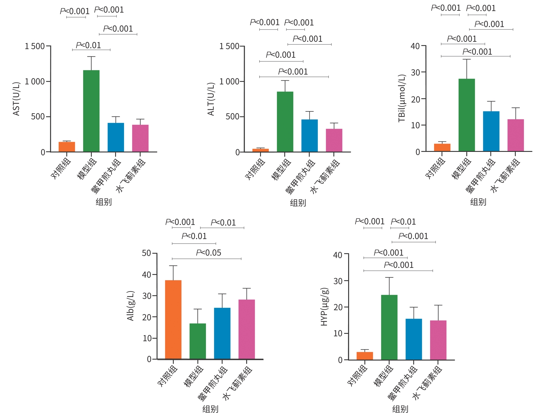
注: AST,天冬氨酸氨基转移酶;ALT,丙氨酸氨基转移酶;TBil,总胆红素;Alb,白蛋白;HYP,羟脯氨酸。
图 2 各组大鼠血清指标检测结果
Figure 2. Detection results of serum parameters in each group of rats
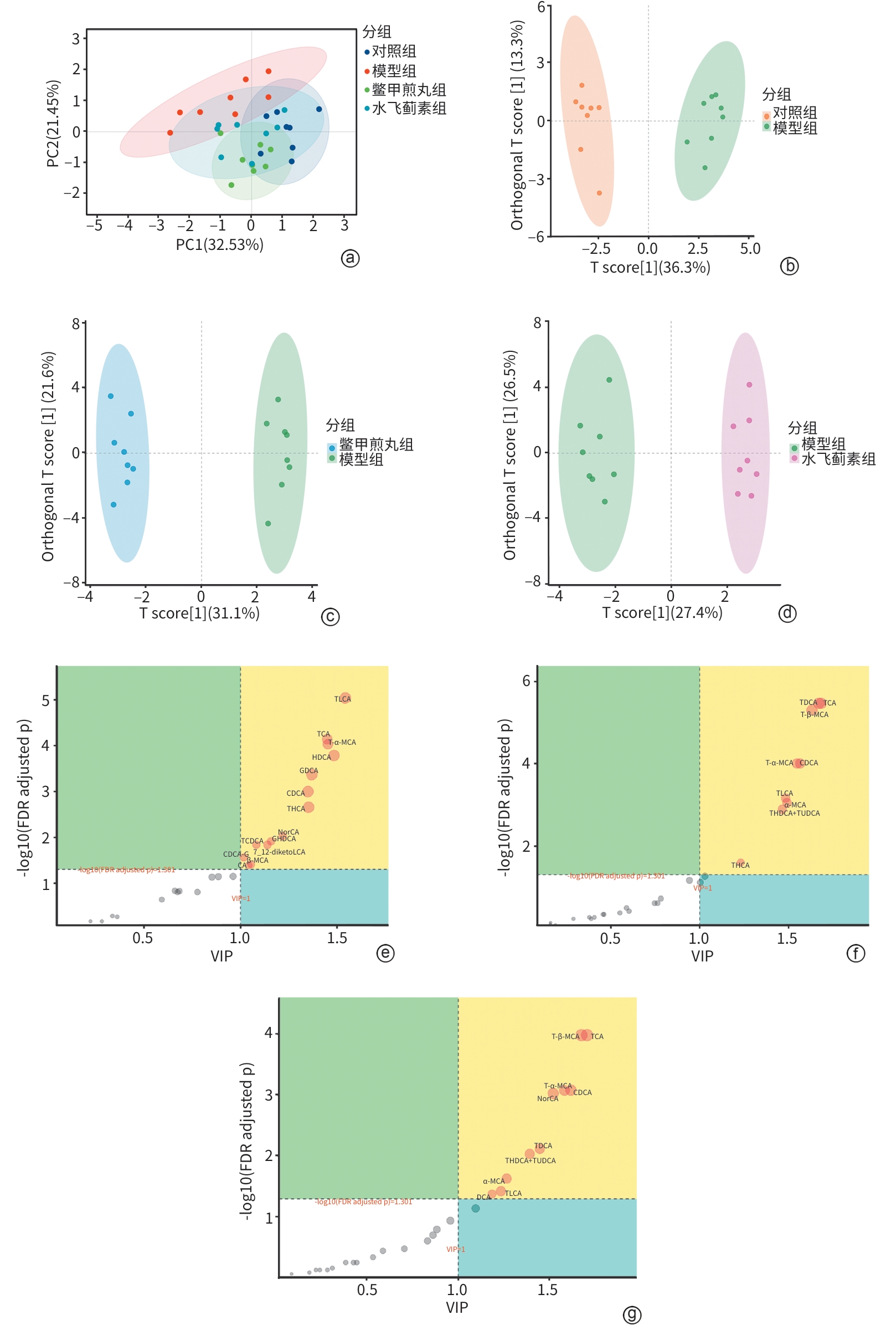
注: a,主成分分析图;b~d,对照组vs模型组、模型组vs鳖甲煎丸组、模型组vs水飞蓟素组的OPLS-DA模型正交信号校正得分图;e~g,对照组vs模型组、模型组vs鳖甲煎丸组、模型组vs水飞蓟素组的OPLS-DA胆汁酸差异图。HDCA,猪去氧胆酸;NorCA,去甲胆酸;β-MCA,β-鼠胆酸;CA,胆酸;TLCA,牛磺石胆酸;T-α-MCA,牛磺-α-鼠胆酸;TCA,牛磺胆酸;GDCA,甘氨脱氧胆酸;THCA,牛磺猪胆酸;CDCA,鹅去氧胆酸;GHDCA,甘氨猪去氧胆酸;7_12-diketoLCA,7,12-二酮石胆酸;TCDCA,牛磺鹅去氧胆酸;CDCA-G,甘氨鹅去氧胆酸;T-β-MCA,牛磺-β-鼠胆酸;THDCA+TUDCA,牛磺猪去氧胆酸+牛磺熊去氧胆酸;α-MCA,α-鼠胆酸;TDCA,牛磺脱氧胆酸;DCA,脱氧胆酸;FDR,假发现率;VIP,变量重要性投影。
图 3 鳖甲煎丸和水飞蓟素调节肝纤维化大鼠的胆汁酸代谢
Figure 3. Biejiajian Pills and silybin regulate bile acid metabolism in rats with liver fibrosis
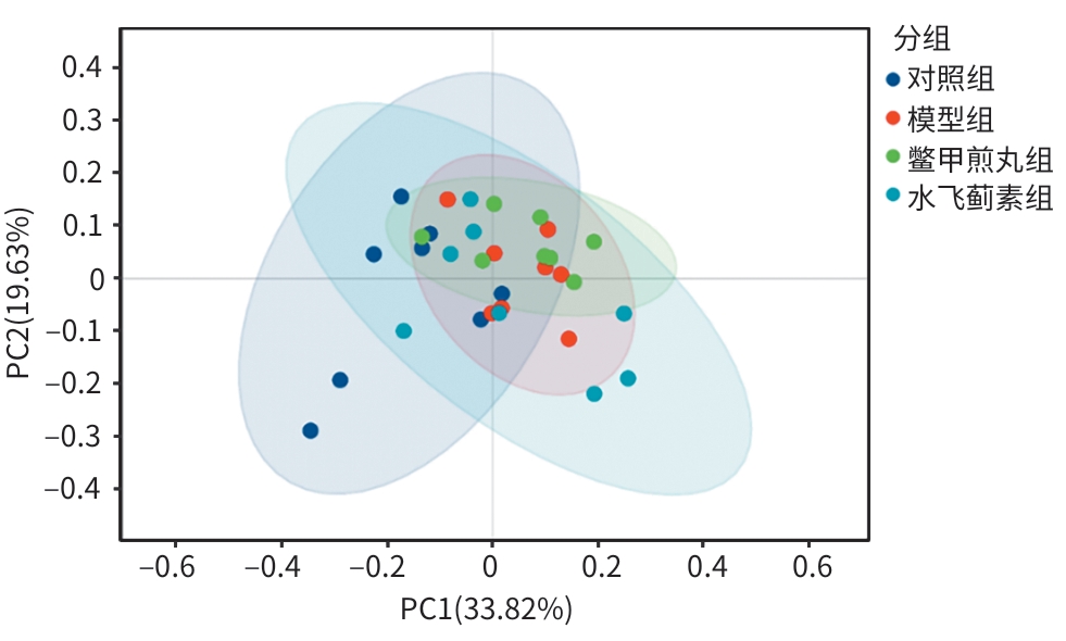
图 4 肝纤维化大鼠模型的短链脂肪酸代谢主成分分析
Figure 4. Principal component analysis diagram of short-chain fatty acid metabolism in rats with liver fibrosis
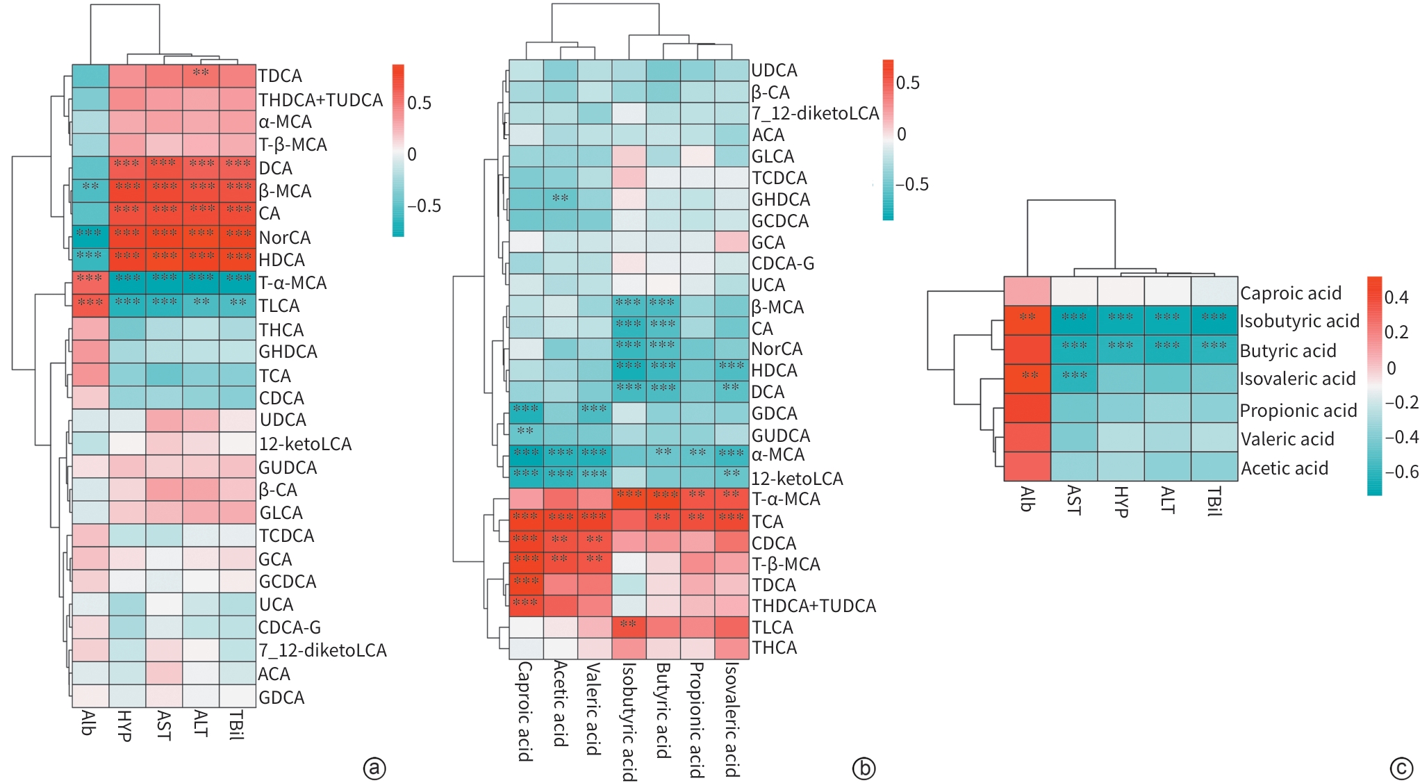
注: a,胆汁酸与血清指标的相关性;b,胆汁酸与短链脂肪酸的相关性;c,短链脂肪酸与血清指标的相关性。TDCA,牛磺脱氧胆酸;THDCA,牛磺猪去氧胆酸;TUDCA,牛磺熊去氧胆酸;α-MCA,α-鼠胆酸;T-β-MCA,牛磺-β-鼠胆酸;DCA,脱氧胆酸;β-MCA,β-鼠胆酸;CA,胆酸;NorCA,去甲胆酸;HDCA,猪去氧胆酸;T-α-MCA,牛磺-α-鼠胆酸;TLCA,牛磺石胆酸;THCA,牛磺猪胆酸;GHDCA,甘氨猪去氧胆酸;TCA,牛磺胆酸;CDCA,鹅去氧胆酸;UDCA,熊去氧胆酸;12-ketoLCA,12-酮石胆酸;GUDCA,甘氨熊去氧胆酸;β-CA,β-胆酸;GLCA,甘氨石胆酸;TCDCA,牛磺鹅去氧胆酸;GCA,甘氨胆酸;GCDCA,甘氨鹅去氧胆酸;UCA,熊胆酸;CDCA-G,甘氨鹅去氧胆酸;7_12-diketoLCA,7,12-二酮石胆酸;ACA,别胆酸;GDCA,甘氨脱氧胆酸;ALT,丙氨酸氨基转移酶;AST,天冬氨酸氨基转移酶;TBil,总胆红素;Alb,白蛋白;HYP,羟脯氨酸;Acetic acid,乙酸;Propionic acid,丙酸;Butyric acid,丁酸;Isobutyric acid,异丁酸;Valeric acid,戊酸;Isovaleric acid,异戊酸;Caproic acid,己酸。*,P<0.05;**,P<0.01;***,P<0.001。
图 5 胆汁酸/短链脂肪酸与血清指标相关性热图
Figure 5. Heatmap showing the correlation between bile acid/short-chain fatty acid and serum indicators
表 1 各组大鼠胆汁酸变化比较
Table 1. Comparison of bile acid changes in rats of each experimental group
组别 动物数
(只)初级胆汁酸
(μg/mL)次级胆汁酸
(μg/mL)未结合型胆汁酸
(μg/mL)甘氨酸结合胆汁酸
(μg/mL)对照组 8 33.69±16.56 7.37±3.43 42.21±19.59 382.01±208.54 模型组 8 57.50±11.84 20.51±4.45 78.01±12.11 474.47±320.09 鳖甲煎丸组 8 34.27±8.031) 13.11±5.301) 47.38±11.021) 237.57±80.83 水飞蓟素组 8 38.34±10.54 13.53±4.751) 51.87±13.131) 360.12±119.75 F值 6.829 11.250 9.845 1.824 P值 0.001 <0.001 0.001 0.166 组别 动物数
(只)牛磺酸结合胆汁酸
(μg/mL)结合型胆汁酸
(μg/mL)总胆汁酸
(μg/mL)对照组 8 3 480.90±647.64 3 862.90±836.07 3 905.11±845.83 模型组 8 2 679.12±724.42 3 153.59±918.34 3 231.60±916.80 鳖甲煎丸组 8 3 756.20±906.041) 3 993.77±960.30 4 041.15±958.94 水飞蓟素组 8 3 650.80±794.311) 4 010.92±752.40 4 062.79±752.10 F值 3.186 1.745 1.617 P值 0.039 0.181 0.208 注:与模型组比较,1)P<0.05。
 下载: 导出CSV
表 2 胆汁酸代谢物的统计分析
Table 2. Statistical analysis of bile acid metabolites
代谢物 模型组vs对照组 模型组vs鳖甲煎丸组 模型组vs水飞蓟素组 P值 VIP P值 VIP P值 VIP 牛磺石胆酸 ↓<0.01 1.52 ↓<0.05 1.48 ↑<0.05 1.22 猪去氧胆酸 ↑<0.01 1.50 — — — — 牛磺-α-鼠胆酸 ↓<0.01 1.47 ↑<0.05 1.54 ↑<0.05 1.52 牛磺胆酸 ↓<0.01 1.46 ↓<0.01 1.64 ↓<0.01 1.60 甘氨脱氧胆酸 ↓<0.01 1.37 — — — — 牛磺猪胆酸 ↓<0.05 1.34 ↓<0.05 1.25 — — 鹅去氧胆酸 ↓<0.05 1.32 ↓<0.01 1.54 ↓<0.01 1.62 去甲胆酸 ↑<0.05 1.20 ↑<0.05 1.03 ↑<0.05 1.48 甘氨猪去氧胆酸 ↓<0.05 1.16 — — — — 7,12-二酮石胆酸 ↓<0.05 1.11 — — — — 牛磺鹅去氧胆酸 ↓<0.05 1.09 — — — — β-鼠胆酸 ↑<0.05 1.06 — — — — 胆酸 ↑<0.05 1.03 — — — — 甘氨鹅去氧胆酸 ↓<0.05 1.02 — — — — 甘氨石胆酸 — — — — — — 熊胆酸 — — — — — — 甘氨胆酸 — — — — ↑<0.05 1.10 脱氧胆酸 — — — — ↑<0.05 1.15 牛磺-β-鼠胆酸 — — ↑<0.01 1.61 ↑<0.01 1.63 牛磺脱氧胆酸 — — ↓<0.01 1.63 ↑<0.01 1.41 熊去氧胆酸 — — — — — — 别胆酸 — — — — — — 牛磺猪去氧胆酸+牛磺熊去氧胆酸 — — ↑<0.05 1.44 ↑<0.01 1.37 α-鼠胆酸 — — ↑<0.05 1.48 ↑<0.01 1.27 β-胆酸 — — — — — — 12-酮石胆酸 — — — — — — 甘氨熊去氧胆酸 — — ↑<0.05 1.00 — — 注:↑,与后组相比上调;↓,与后组相比下调。VIP,变量重要性投影;VIP>1,对两组间的差异有显著贡献;VIP<1,对分组的贡献较小。—,P>0.05或VIP<1。
下载: 导出CSV
表 3 各实验组大鼠短链脂肪酸变化比较
Table 3. Comparison of short-chain fatty acid changes in rats of each experimental group
组别 动物数
(只)乙酸
(μg/g)丙酸
(μg/g)异丁酸
(μg/g)丁酸
(μg/g)异戊酸
(μg/g)戊酸
(μg/g)己酸
(μg/g)对照组 8 1 388.22±286.45 517.23±156.46 52.50±13.42 310.87±61.73 32.33±7.23 61.04±9.77 1.81±0.07 模型组 8 1 065.74±296.86 327.52±57.69 26.19±3.65 158.84±24.40 24.79±4.88 46.01±5.76 1.36±0.42 鳖甲煎丸组 8 1 648.83±362.531) 500.07±68.011) 43.57±4.261) 286.35±54.631) 34.25±10.081) 79.57±9.201) 3.57±0.221) 水飞蓟素组 8 1 527.84±343.871) 508.52±90.001) 42.15±4.46 269.47±49.40 33.86±7.27 70.46±13.271) 2.49±0.341) F值 4.824 6.511 16.560 14.710 2.717 16.870 85.780 P值 0.008 0.002 <0.001 <0.001 0.047 <0.001 <0.001 注:与模型组比较,1)P<0.05。
下载: 导出CSV
-
[1] Roehlen N, Crouchet E, Baumert T F. Liver fibrosis: Mechanistic concepts and therapeutic perspectives[J]. Cells, 2020, 9( 4): 875. DOI: 10.3390/cells9040875. [2] Li H. Angiogenesis in the progression from liver fibrosis to cirrhosis and hepatocelluar carcinoma[J]. Expert Rev Gastroenterol Hepatol, 2021, 15( 3): 217- 233. DOI: 10.1080/17474124.2021.1842732. [3] de Franchis R, Bosch J, Garcia-Tsao G, et al. Baveno VII-Renewing consensus in portal hypertension[J]. J Hepatol, 2022, 76( 4): 959- 974. DOI: 10.1016/j.jhep.2021.12.022. [4] Arroyo V, Moreau R, Jalan R, et al. Acute-on-chronic liver failure: A new syndrome that will re-classify cirrhosis[J]. J Hepatol, 2015, 62( 1): S131- S143. DOI: 10.1016/j.jhep.2014.11.045. [5] Vilstrup H, Amodio P, Bajaj J, et al. Hepatic encephalopathy in chronic liver disease: 2014 Practice Guideline by the American Association for the Study of Liver Diseases and the European Association for the Study of the Liver[J]. Hepatology, 2014, 60( 2): 715- 735. DOI: 10.1002/hep.27210. [6] Llovet J M, Kelley R K, Villanueva A, et al. Hepatocellular carcinoma[J]. Nat Rev Dis Primers, 2021, 7( 1): 6. DOI: 10.1038/s41572-020-00240-3. [7] Liu Z P, Ouyang G Q, Huang G Z, et al. Global burden of cirrhosis and other chronic liver diseases due to nonalcoholic fatty liver disease, 1990-2019[J]. World J Hepatol, 2023, 15( 11): 1210- 1225. DOI: 10.4254/wjh.v15.i11.1210. [8] Parada Venegas D, de la Fuente M K, Landskron G, et al. Corrigendum: Short chain fatty acids(SCFAs)-mediated gut epithelial and immune regulation and its relevance for inflammatory bowel diseases[J]. Front Immunol, 2019, 10: 1486. DOI: 10.3389/fimmu.2019.01486. [9] Chiang J Y L, Ferrell J M. Bile acid metabolism in liver pathobiology[J]. Gene Expr, 2018, 18( 2): 71- 87. DOI: 10.3727/105221618X15156018385515. [10] Liu Sihong, Li Shasha, Hou Youjuan, et al. Ancient and modern literature on clinical application of Biejia Jianwan in medical treasures of the golden chamber[J]. Chin J Exp Tradit Med Formulae, 2020, 26( 6): 12- 17. DOI: 10.13422/j.cnki.syfjx.20192321.刘思鸿, 李莎莎, 侯酉娟, 等.《金匮要略》鳖甲煎丸临床应用的古今文献研究[J]. 中国实验方剂学杂志, 2020, 26( 6): 12- 17. DOI: 10.13422/j.cnki.syfjx.20192321. [11] Luo Weikang, Wang Xianbei, Wang Jiafeng. Understanding of the synopsis of turtle-fried pill in golden chamber[J]. J Sichuan Tradit Chin Med, 2018, 36( 4): 25- 28.罗伟康, 王宪贝, 王加锋. 浅谈对《金匮要略》鳖甲煎丸方证的理解[J]. 四川中医, 2018, 36( 4): 25- 28. [12] Sun Haitao, Wen Bin, Chen Guanxin, et al. Effect of Biejiajian pill on Wnt/β-catenin pathway associated protein and its target gene expression in liver tissue of hepatic fibrosis model rats[J]. J Tradit Chin Med, 2018, 59( 10): 876- 881. DOI: 10.13288/j.11-2166/r.2018.10.015.孙海涛, 文彬, 陈冠新, 等. 鳖甲煎丸对肝纤维化模型大鼠肝组织中Wnt/β-catenin信号通路相关蛋白及其靶基因表达的影响[J]. 中医杂志, 2018, 59( 10): 876- 881. DOI: 10.13288/j.11-2166/r.2018.10.015. [13] Bataller R, Brenner D A. Liver fibrosis[J]. J Clin Invest, 2005, 115( 2): 209- 218. DOI: 10.1172/jci24282. [14] Albillos A, de Gottardi A, Rescigno M. The gut-liver axis in liver disease: Pathophysiological basis for therapy[J]. J Hepatol, 2020, 72( 3): 558- 577. DOI: 10.1016/j.jhep.2019.10.003. [15] Liu Yunxiao, Dou Jing, Wang Xiaozhong. Association between liver fibrosis and gut microbiota based on“harmonizing liver and spleen”[J]. J Clin Hepatol, 2023, 39( 2): 278- 283. DOI: 10.3969/j.issn.1001-5256.2023.02.005.刘云霄, 窦婧, 王晓忠. 基于“调和肝脾”论述肝纤维化与肠道菌群的关系[J]. 临床肝胆病杂志, 2023, 39( 2): 278- 283. DOI: 10.3969/j.issn.1001-5256.2023.02.005. [16] Wiest R, Albillos A, Trauner M, et al. Targeting the gut-liver axis in liver disease[J]. J Hepatol, 2017, 67( 5): 1084- 1103. DOI: 10.1016/j.jhep.2017.05.007. [17] Koh A, de Vadder F, Kovatcheva-Datchary P, et al. From dietary fiber to host physiology: Short-chain fatty acids as key bacterial metabolites[J]. Cell, 2016, 165( 6): 1332- 1345. DOI: 10.1016/j.cell.2016.05.041. [18] Seki E, Schwabe R F. Hepatic inflammation and fibrosis: Functional links and key pathways[J]. Hepatology, 2015, 61( 3): 1066- 1079. DOI: 10.1002/hep.27332. [19] Chen Bin, Xu Jiawei, Peng Jie, et al. Effect of“treating liver by nourishing spleen” on gut microbiota in rats with liver fibrosis based on Xiaoyao powder and its separated recipe[J]. J Clin Hepatol, 2016, 32( 4): 657- 662. DOI: 10.3969/j.issn.1001-5256.2016.04.009.陈斌, 徐嘉蔚, 彭杰, 等. 基于逍遥散及其拆方研究“肝病实脾法”对肝纤维化大鼠肠道菌群的影响[J]. 临床肝胆病杂志, 2016, 32( 4): 657- 662. DOI: 10.3969/j.issn.1001-5256.2016.04.009. [20] Kremer A E, Mayo M J, Hirschfield G M, et al. Seladelpar treatment reduces IL-31 and pruritus in patients with primary biliary cholangitis[J]. Hepatology, 2024, 80( 1): 27- 37. DOI: 10.1097/HEP.0000000000000728. [21] Peng L Y, Li Z R, Green R S, et al. Butyrate enhances the intestinal barrier by facilitating tight junction assembly via activation of AMP-activated protein kinase in Caco-2 cell monolayers[J]. J Nutr, 2009, 139( 9): 1619- 1625. DOI: 10.3945/jn.109.104638. [22] Xu Q, Zhang R S, Mu Y, et al. Propionate ameliorates alcohol-induced liver injury in mice via the gut-liver axis: Focus on the improvement of intestinal permeability[J]. J Agric Food Chem, 2022, 70( 20): 6084- 6096. DOI: 10.1021/acs.jafc.2c00633. [23] Zhang L, Chen J, Ge S J, et al. Propionic acid secreted by Akkermansia muciniphila alleviates hepatic fibrosis by antioxidant regulation across the gut-liver axis[J]. Life Metab, 2026, 5( 1): loaf036. DOI: 10.1093/lifemeta/loaf036. [24] Zhang C Y, Liu S, Sui Y X, et al. Roles of short-chain fatty acids in metabolic dysfunction-associated steatotic liver disease and metabolic dysfunction-associated steatohepatitis[J]. World J Hepatol, 2025, 17( 11): 113756. DOI: 10.4254/wjh.v17.i11.113756. -

 本文二维码
本文二维码
计量
- 文章访问数: 18
- HTML全文浏览量: 8
- PDF下载量: 1
- 被引次数: 0


