
 PDF下载 ( 7583 KB)

慢性乙型肝炎儿童肝组织HBsAg和HBcAg表达特征谱及其与血清学标志物的相关性分析
DOI: 10.12449/JCH251013
Expression profiles of HBsAg and HBcAg in liver tissue and their correlation with serological markers in children with chronic hepatitis B
-
摘要:
目的 分析慢性乙型肝炎患儿肝组织内HBsAg和HBcAg表达特点,并评估其与血清HBV标志物的关联。 方法 连续纳入2013年1月—2023年12月中国人民解放军总医院第五医学中心257例肝穿刺确诊CHB患者。采用NIS-Elements系统采集肝组织HBsAg和HBcAg免疫组化图像,Image J软件定量分析。计数资料组内比较采用单样本χ2检验,并通过Pearson/Spearman/Kendall’s Tau-b相关分析评估病毒抗原表达与血清学标志物的相关性。 结果 257例CHB患者中儿童162例(<5岁76例,5~18岁86例),成人95例。患者肝组织内HBsAg表达模式、面积、强度和HBcAg表达面积、强度分布在不同年龄组及儿童不同HBeAg状态均存在显著差异(P值均<0.05)。<5岁组中,HBsAg染色面积与抗-HBs、HBeAg呈显著负相关(P值均<0.05),与ALT、AST呈显著正相关(P值均<0.05);HBsAg染色强度与qHBsAg呈显著正相关(P<0.05),与抗-HBs呈显著负相关(P<0.05)。儿童组中,HBsAg染色面积与抗-HBs、HBeAg均呈负相关(P值均<0.05);HBsAg染色强度与qHBsAg呈正相关(P<0.05),与抗-HBs呈负相关(P<0.05)。成人组中,HBsAg染色面积与ALT、AST和肝组织炎症活动度均呈正相关(P值均<0.05);HBsAg染色强度与qHBsAg、HBeAg、HBV DNA均呈正相关(P值均<0.05),与肝脏炎症活动度和肝纤维化程度均呈负相关(P值均<0.05)。在<5岁组中,HBcAg的染色面积与qHBsAg、HBV DNA水平呈正相关(P值均<0.05);HBcAg的染色强度与HBV DNA呈显著正相关(P<0.001)。在5~18岁组中,HBcAg的染色面积和强度与qHBsAg、HBeAg及HBV DNA水平均呈正相关(P值均<0.05)。儿童组中,HBcAg的染色面积与qHBsAg、HBeAg及HBV DNA水平呈正相关(P值均<0.05);HBcAg的染色强度与qHBsAg、HBV DNA水平呈正相关(P值均<0.05)。成人组中,HBcAg的染色面积和强度与qHBsAg、HBeAg及HBV DNA水平均呈显著正相关(P值均<0.001),HBcAg的染色面积与血清中ALT水平也存在正相关性(P=0.043)。 结论 CHB儿童患者肝内HBsAg和HBcAg表达与血清学标志物具有良好的相关性,在临床实践中,可结合血清学指标评估儿童CHB患者的肝脏状况,确定免疫分期,并为治疗时机选择提供循证依据。 Abstract:Objective To investigate the expression features of HBsAg and HBcAg in liver tissue and their correlation with HBV serum markers in children with chronic hepatitis B (CHB). Methods A total of 257 patients who were consecutively admitted to The Fifth Medical Center of Chinese PLA General Hospital from January 2013 to December 2023 and underwent liver biopsy to achieve a confirmed diagnosis of CHB were enrolled in this study. The NIS-Elements system was used to capture the immunohistochemical images of HBsAg and HBcAg in liver tissues, and Image J software was used for quantitative analysis. The one-sample chi-square test was used for within-group comparison of continuous data, and the Pearson/Spearman/Kendall’s Tau-b correlation analysis was used to investigate the correlation between viral antigen expression and serological markers. Results Among the 257 CHB patients, there were 162 children (76 children aged<5 years and 86 children aged 5 — 18 years) and 95 adults. There were significant differences in the expression pattern, area, and intensity of HBsAg and the area and intensity of HBcAg in liver tissue between different age groups and between the children with different HBeAg statuses (all P<0.05). In the children aged<5 years, HBsAg staining area was significantly negatively correlated with anti-HBs and HBeAg (both P<0.05)and was significantly positively correlated with ALT and AST (both P<0.05), and HBsAg staining intensity was significantly positively correlated with qHBsAg (P<0.05) and was significantly negatively correlated with anti-HBs (P<0.05). In the children group, HBsAg staining area was negatively correlated with anti-HBs and HBeAg (both P<0.05), and HBsAg staining intensity was positively correlated with qHBsAg (P<0.05) and was negatively correlated with anti-HBs (P<0.05). In the adult group, HBsAg staining area was positively correlated with ALT, AST, and liver inflammatory activity (all P<0.05), and HBsAg staining intensity was positively correlated with qHBsAg, HBeAg, and HBV DNA (all P<0.05) and was negatively correlated with liver inflammatory activity and fibrosis degree (both P<0.05). In the children aged<5 years, HBcAg staining area was positively correlated with qHBsAg and HBV DNA (both P<0.05), and HBcAg staining intensity was significantly positively correlated with HBV DNA (P<0.001). In the children aged 5 — 18 years, the area and intensity of HBcAg staining were positively correlated with qHBsAg, HBeAg, and HBV DNA (all P<0.05). In the children group, HBcAg staining area was positively correlated with qHBsAg, HBeAg, and HBV DNA (all P<0.05), and HBcAg staining intensity was positively correlated with qHBsAg and HBV DNA (both P<0.05). In the adult group, the area and intensity of HBcAg staining were positively correlated with qHBsAg, HBeAg, and HBV DNA (all P<0.001), and HBcAg staining area was positively correlated with the serum level of ALT (P=0.043). Conclusion The expression levels of HBsAg and HBcAg in liver tissue of children with CHB are significantly correlated with serological markers, and in clinical practice, HBsAg and HBcAg combined with serological markers can help to assess the condition of the liver, determine the immune stage, and provide evidence-based guidance for treatment timing. -
Key words:
- Hepatitis B, Chronic /
- Hepatitis B Surface Antigens /
- Hepatitis B Core Antigens /
- Biomarkers /
- Child
-
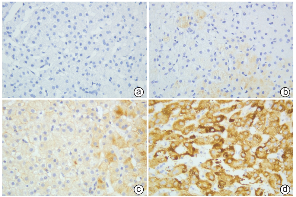
注: a,无染色(阴性);b,浅黄色;c,棕黄色;d,深褐色。
图 1 免疫组化染色检测HBsAg的染色着色情况(×400)
Figure 1. Detection of hepatitis B antigen staining by immunohistochemical staining
表 1 257例CHB患者病毒学、血清学和组织学特征
Table 1. Virological, serobiochemical, and histological characteristics
项目 数值 qHBsAg(log10 IU/mL) 3.96(3.33~4.50) 抗-HBs(log10 COI) 0.30(0.30~0.30) HBeAg(log10 COI) 2.70(-0.91~3.14) HBeAg阳性[例(%)] 175(68.09) HBV DNA(log10 IU/mL) 6.89(3.74~8.00) HBV基因型[例(%)] B 38(14.79) C 144(56.03) D 3(1.17) 未检测出 72(28.02) ALT(U/L) 84.00(38.00~173.00) ALT升高[例(%)] 195(75.88) AST(U/L) 67.00(40.00~126.00) TBil(μmol/L) 8.90(6.35~12.10) Alb(g/L) 40.00(38.00~43.00) 肝组织炎症活动度[例(%)] G0~G1 97(37.74) G2~G3 160(62.26) 肝纤维化程度[例(%)] S0~S1 119(46.30) S2~S3 138(53.70) 免疫组化[例(%)] HBsAg阳性 235(91.44) HBcAg阳性 150(58.37)  下载: 导出CSV
表 2 HBsAg表达模式差异性
Table 2. Heterogeneity in HBsAg expression patterns
项目 例数 表达模式[例(%)] χ2值 P值 阴性 胞浆着色 胞膜、胞浆着色 儿童组 162 13(8.02) 77(47.53) 72(44.44) 46.926 <0.001 <5岁 76 7(9.21) 40(52.63) 29(38.16) 22.289 <0.001 5~18岁 86 6(6.98) 37(43.02) 43(50.00) 27.512 <0.001 成人组 95 9(9.47) 33(34.74) 53(55.79) 30.653 <0.001 儿童组HBeAg状态 阴性 52 4(7.69) 25(48.08) 23(44.23) 15.500 <0.001 阳性 110 9(8.18) 52(47.27) 49(44.55) 31.436 <0.001
下载: 导出CSV
表 3 HBsAg表达面积差异性
Table 3. Heterogeneity in HBsAg expression areas
项目 例数 表达面积[例(%)] χ2值 P值 0 <5% 5%~20% >20% 儿童组 162 13(8.02) 62(38.27) 32(19.75) 55(33.95) 37.062 <0.001 <5岁 76 7(9.21) 34(44.74) 12(15.79) 23(30.26) 22.842 <0.001 5~18岁 86 6(6.98) 28(32.56) 23(23.26) 32(37.21) 18.372 <0.001 成人组 95 9(9.47) 26(27.37) 31(32.63) 29(30.53) 12.747 <0.001 儿童组HBeAg状态 阴性 52 4(7.69) 14(26.92) 13(25.00) 21(40.38) 11.231 0.011 阳性 110 9(8.18) 48(43.64) 19(17.27) 34(30.91) 31.891 <0.001
下载: 导出CSV
表 4 HBsAg表达强度差异性
Table 4. Heterogeneity in HBsAg expression intensities
项目 例数 表达强度[例(%)] χ2值 P值 无染色 浅黄色 棕黄色 深褐色 儿童组 162 13(8.02) 32(19.75) 60(37.04) 57(35.19) 36.568 <0.001 <5岁 76 7(9.21) 18(23.68) 23(30.26) 28(36.84) 12.737 0.005 5~18岁 86 6(6.98) 14(16.28) 37(43.02) 29(33.72) 27.581 <0.001 成人组 95 9(9.47) 5(5.26) 33(34.74) 48(50.53) 52.326 <0.001 儿童组HBeAg状态 阴性 52 4(7.69) 8(15.38) 19(36.54) 21(40.38) 15.846 0.001 阳性 110 9(8.18) 24(21.82) 41(37.27) 36(32.73) 22.145 <0.001
下载: 导出CSV
表 5 儿童和成人患者HBsAg染色结果与血清学及组织学指标的相关性
Table 5. Correlation between HBsAg staining results and serological and histological indicators in pediatric and adult patients
项目 HBsAg染色 <5岁组(n=76) 5~18岁组(n=86) 儿童组(n=162) 成人组(n=95) r值 P值 r值 P值 r值 P值 r值 P值 qHBsAg 面积 0.072 0.536 0.141 0.194 0.094 0.234 0.198 0.055 强度 0.246 0.032 0.133 0.222 0.183 0.020 0.322 0.001 抗-HBs 面积 -0.308 0.007 -0.103 0.345 -0.203 0.010 -0.149 0.644 强度 -0.351 0.002 -0.080 0.464 -0.218 0.005 -0.495 0.102 HBeAg 面积 -0.325 0.004 -0.139 0.201 -0.234 0.003 0.060 0.561 强度 -0.039 0.739 0.013 0.903 -0.023 0.772 0.212 0.039 HBV DNA 面积 0.108 0.352 0.014 0.901 0.069 0.383 0.186 0.071 强度 0.117 0.313 0.147 0.177 0.127 0.108 0.208 0.043 ALT 面积 0.251 0.029 0.040 0.712 0.136 0.085 0.274 0.007 强度 -0.064 0.580 -0.126 0.247 -0.105 0.182 0.057 0.585 AST 面积 0.314 0.006 0.054 0.618 0.145 0.067 0.221 0.032 强度 -0.015 0.895 -0.206 0.057 -0.121 0.125 -0.110 0.290 TBil 面积 0.171 0.139 -0.056 0.606 0.081 0.307 -0.107 0.303 强度 0.113 0.332 0.014 0.899 0.065 0.415 -0.127 0.221 Alb 面积 -0.011 0.925 0.104 0.342 0.026 0.747 0.108 0.297 强度 -0.114 0.327 -0.003 0.975 -0.065 0.415 0.089 0.389 肝组织炎症活动度 面积 0.122 0.227 0.033 0.749 0.069 0.346 0.178 0.034 强度 0.097 0.363 -0.075 0.476 0.007 0.923 -0.235 0.007 肝纤维化程度 面积 0.179 0.085 0.021 0.837 0.090 0.216 0.060 0.531 强度 0.097 0.375 -0.121 0.209 -0.018 0.799 -0.214 0.014
下载: 导出CSV
表 6 HBcAg表达模式差异性
Table 6. Heterogeneity in HBcAg expression patterns
项目 例数 表达模式[例(%)] χ2值 P值 阴性 胞浆着色 胞膜、胞浆着色 儿童组 162 67(41.36) 52(32.10) 43(26.54) 5.444 0.066 <5岁 76 34(44.74) 24(31.58) 18(23.68) 5.158 0.076 5~18岁 86 33(38.37) 28(32.56) 25(29.07) 1.140 0.566 成人组 95 40(42.11) 25(26.32) 30(31.58) 3.684 0.158 儿童组HBeAg状态 阴性 52 29(55.77) 20(38.46) 3(5.77) 20.115 <0.001 阳性 110 38(34.55) 32(29.09) 40(36.36) 0.945 0.623
下载: 导出CSV
表 7 HBcAg表达面积差异性
Table 7. Heterogeneity in HBcAg expression areas
项目 例数 表达面积[例(%)] χ2值 P值 0 <5% 5%~20% >20% 儿童组 162 67(41.36) 33(20.37) 51(31.48) 11(6.79) 42.938 <0.001 <5岁 76 34(44.74) 15(19.74) 22(28.95) 5(6.58) 23.474 <0.001 5~18岁 86 33(38.37) 18(20.93) 29(33.72) 6(6.98) 20.512 <0.001 成人组 95 40(42.11) 17(17.89) 26(27.37) 12(12.63) 19.063 <0.001 儿童组HBeAg状态 阴性 52 29(55.77) 8(15.38) 13(25.00) 2(3.85) 30.923 <0.001 阳性 110 38(34.55) 25(22.73) 38(34.55) 9(8.18) 20.691 <0.001
下载: 导出CSV
表 8 HBcAg表达强度差异性
Table 8. Heterogeneity in HBcAg expression intensities
项目 例数 表达强度[例(%)] χ2值 P值 无染色 浅黄色 棕黄色 深褐色 儿童组 162 67(41.36) 43(26.54) 43(26.54) 9(5.56) 42.148 <0.001 <5岁 76 34(44.74) 23(30.26) 16(21.05) 3(3.95) 26.632 0.005 5~18岁 86 33(38.37) 20(23.26) 27(31.40) 6(6.98) 18.837 <0.001 成人组 95 40(42.11) 13(13.68) 27(28.42) 15(15.79) 19.653 <0.001 儿童组HBeAg状态 阴性 52 29(55.77) 6(11.54) 16(30.77) 1(1.92) 35.231 <0.001 阳性 110 38(34.55) 37(33.64) 27(24.55) 8(7.27) 21.127 <0.001
下载: 导出CSV
表 9 儿童和成人患者HBcAg染色结果与血清学及组织学指标的相关性
Table 9. Correlation between HBcAg staining results and serological and histological indicators in pediatric and adult patients
项目 HBcAg染色 <5岁组(n=76) 5~18岁组(n=86) 儿童组(n=162) 成人组(n=95) r值 P值 r值 P值 r值 P值 r值 P值 qHBsAg 面积 0.288 0.012 0.273 0.011 0.274 <0.001 0.619 <0.001 强度 0.211 0.067 0.277 0.010 0.228 0.003 0.588 <0.001 抗-HBs 面积 -0.035 0.763 0.036 0.742 -0.006 0.942 -0.234 0.465 强度 -0.039 0.737 0.032 0.771 -0.013 0.873 -0.367 0.240 HBeAg 面积 -0.053 0.649 0.401 <0.001 0.219 0.005 0.701 <0.001 强度 -0.010 0.928 0.283 0.008 0.130 0.099 0.629 <0.001 HBV DNA 面积 0.580 <0.001 0.409 <0.001 0.499 <0.001 0.567 <0.001 强度 0.554 <0.001 0.331 0.002 0.449 <0.001 0.546 <0.001 ALT 面积 0.029 0.802 0.023 0.834 0.024 0.762 0.208 0.043 强度 0.064 0.585 -0.022 0.837 0.020 0.804 0.197 0.056 AST 面积 -0.092 0.430 0.027 0.803 -0.023 0.774 0.107 0.303 强度 -0.044 0.708 -0.033 0.761 -0.036 0.651 0.114 0.272 TBil 面积 0.040 0.734 -0.063 0.565 0.001 0.992 -0.138 0.184 强度 0.118 0.309 -0.126 0.247 0.017 0.828 -0.160 0.121 Alb 面积 0.067 0.567 0.145 0.182 0.088 0.266 -0.110 0.290 强度 0.091 0.435 0.090 0.410 0.060 0.450 -0.052 0.614 肝组织炎症活动度 面积 -0.134 0.229 0.100 0.316 -0.010 0.895 -0.001 0.991 强度 -0.111 0.316 0.124 0.221 0.020 0.789 -0.024 0.789 肝纤维化程度 面积 -0.086 0.401 -0.192 0.060 -0.140 0.054 -0.138 0.150 强度 -0.069 0.510 -0.083 0.418 -0.071 0.331 -0.147 0.099
下载: 导出CSV
-
[1] BURKI T. WHO’s 2024 global hepatitis report[J]. Lancet Infect Dis, 2024, 24( 6): e362- e363. DOI: 10.1016/S1473-3099(24)00307-4. [2] INDOLFI G, EASTERBROOK P, DUSHEIKO G, et al. Hepatitis B virus infection in children and adolescents[J]. Lancet Gastroenterol Hepatol, 2019, 4( 6): 466- 476. DOI: 10.1016/S2468-1253(19)30042-1. [3] JENG WJ, PAPATHEODORIDIS GV, LOK ASF. Hepatitis B[J]. Lancet, 2023, 401( 10381): 1039- 1052. DOI: 10.1016/S0140-6736(22)01468-4. [4] Chinese Society of Infectious Diseases, Chinese Medical Association; Chinese Society of Hepatology, Chinese Medical Association; Group of Infectious Diseases, Chinese Pediatric Society, Chinese Medical Association, et al. Expert consensus on the prevention and treatment of chronic hepatitis B in children[J]. Infect Dis Info, 2024, 37( 2): 97- 112. DOI: 10.3969/j.issn.1007-8134.2024.02.001.中华医学会感染病学分会, 中华医学会肝病学分会, 中华医学会儿科学分会感染学组, 等. 儿童慢性乙型肝炎防治专家共识[J]. 传染病信息, 2024, 37( 2): 97- 112. DOI: 10.3969/j.issn.1007-8134.2024.02.001. [5] SAFAIE P, POONGKUNRAN M, KUANG PP, et al. Intrahepatic distribution of hepatitis B virus antigens in patients with and without hepatocellular carcinoma[J]. World J Gastroenterol, 2016, 22( 12): 3404- 3411. DOI: 10.3748/wjg.v22.i12.3404. [6] ALPSOY A, ADANIR H, BAYRAMOGLU Z, et al. Correlation of hepatitis B surface antigen expression with clinicopathological and biochemical parameters in liver biopsies: A comprehensive study[J]. World J Hepatol, 2022, 14( 1): 260- 273. DOI: 10.4254/wjh.v14.i1.260. [7] ZHANG XM, MA XQ, XU ZJ, et al. Effect of drug-resistant mutations in reverse transcriptase region of hepatitis B virus on the level of serum hepatitis B surface antigen[J/CD]. Chin J Exp Clin Infect Dis(Electronic Edition), 2023, 17( 5): 324- 332. DOI: 10.3877/cma.j.issn.1674-1358.2023.05.006.张小曼, 马筱秋, 许正锯, 等. 乙型肝炎病毒逆转录酶区耐药突变对血清乙型肝炎病毒表面抗原水平的影响[J/CD]. 中华实验和临床感染病杂志(电子版), 2023, 17( 5): 324- 332. DOI: 10.3877/cma.j.issn.1674-1358.2023.05.006. [8] Chinese Society of Hepatology and Chinese Society of Infectious Diseases, Chinese Medical Association. The guideline of prevention and treatment for chronic hepatitis B(2010 version)[J]. J Clin Hepatol, 2011, 27( 1): 113- 128.中华医学会肝病学分会, 中华医学会感染病学分会. 慢性乙型肝炎防治指南(2010年版)[J]. 临床肝胆病杂志, 2011, 27( 1): 113- 128. [9] National Health Commission. Reference intervals of clinical biochemistry tests commonly used for children: WS/T 780-2021[S/OL].( 2021-04-09). http://www.nhc.gov.cn/wjw/s9492/202105/170ca00246014d18b82a61cabf9fdb2f.shtml. http://www.nhc.gov.cn/wjw/s9492/202105/170ca00246014d18b82a61cabf9fdb2f.shtml国家卫生健康委员会. 儿童临床常用生化检验项目参考区间: WS/T 780-2021[S/OL].( 2021-04-09). http://www.nhc.gov.cn/wjw/s9492/202105/170ca00246014d18b82a61cabf9fdb2f.shtml. http://www.nhc.gov.cn/wjw/s9492/202105/170ca00246014d18b82a61cabf9fdb2f.shtml [10] SCHEUER PJ. Classification of chronic viral hepatitis: A need for reassessment[J]. J Hepatol, 1991, 13( 3): 372- 374. DOI: 10.1016/0168-8278(91)90084-o. [11] Chinese Society of Hepatology, Chinese Medical Association; Chinese Society of Infectious Diseases, Chinese Medical Association. Guidelines for the prevention and treatment of chronic hepatitis B(version 2022)[J]. Infect Dis Info, 2023, 36( 1): 1- 17. DOI: 10.3969/j.issn.1007-8134.2023.01.01.中华医学会肝病学分会, 中华医学会感染病学分会. 慢性乙型肝炎防治指南(2022年版)[J]. 传染病信息, 2023, 36( 1): 1- 17. DOI: 10.3969/j.issn.1007-8134.2023.01.01. [12] SU TH, LIU CJ, YANG HC, et al. Clinical significance and evolution of hepatic HBsAg expression in HBeAg-positive patients receiving interferon therapy[J]. J Gastroenterol, 2014, 49( 2): 356- 362. DOI: 10.1007/s00535-013-0840-z. [13] LIU L, LIU CY, LI JY, et al. Analysis of pathological changes of liver tissue in patients with low level HBsAg[J]. Chongqing Med, 2018, 47( 8): 1109- 1111. DOI: 10.3969/j.issn.1671-8348.2018.08.032.刘立, 刘春云, 李俊义, 等. 低水平HBsAg患者肝脏组织病理变化分析[J]. 重庆医学, 2018, 47( 8): 1109- 1111. DOI: 10.3969/j.issn.1671-8348.2018.08.032. [14] RAMAKRISHNA B, MUKHOPADHYA A, KURIAN G. Correlation of hepatocyte expression of hepatitis B viral antigens with histological activity and viral titer in chronic hepatitis B virus infection: An immunohistochemical study[J]. J Gastroenterol Hepatol, 2008, 23( 11): 1734- 1738. DOI: 10.1111/j.1440-1746.2008.05416.x. [15] SERINOZ E, VARLI M, ERDEN E, et al. Nuclear localization of hepatitis B core antigen and its relations to liver injury, hepatocyte proliferation, and viral load[J]. J Clin Gastroenterol, 2003, 36( 3): 269- 272. DOI: 10.1097/00004836-200303000-00016. [16] UZUN Y, BOZKAYA H, ERDEN E, et al. Hepatitis B core antigen expression pattern reflects the response to anti-viral treatment[J]. J Gastroenterol Hepatol, 2006, 21( 6): 977- 981. DOI: 10.1111/j.1440-1746.2006.04263.x. [17] GIADANS CG, RÍOS DA, AMEIGEIRAS B, et al. Chronic hepatitis B: The interplay between intrahepatic lymphocyte population and viral antigens in relation to liver damage[J]. J Viral Hepat, 2019, 26( 6): 727- 737. DOI: 10.1111/jvh.13078. -

 本文二维码
本文二维码
计量
- 文章访问数: 391
- HTML全文浏览量: 111
- PDF下载量: 101
- 被引次数: 0

